Dr. A.K. Singal MBBS (Gold Medalist), DNB, MNAMS
M.CH (AIIMS, Gold Medalist)
Stecker Scholar Pediatric Urology (USA)
M.CH (AIIMS, Gold Medalist)
Stecker Scholar Pediatric Urology (USA)
Spina bifida (myelomeningocele) is a condition in which the lower part of the spinal cord does not form normally. Infants with this condition are born with remnants of the abnormal spinal cord enclosed in a sac on their back. These infants can have a number of problems, but this article will focus on problems in their urinary tract due to meningomyelocele also called neuropathic or neurogenic bladder.
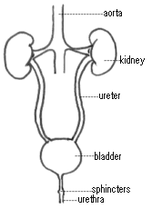
The urinary tract consists of the kidneys, ureters, bladder and urethra. The kidneys are the organs that are responsible for filtering waste products from the bloodstream and produce urine continuously. The urine drains down tubes called ureters to the bladder, which normally stores urine and empties intermittently by muscular contraction. The urine exits the bladder through the urethra in a process is called voiding or urination. Sphincters are special muscles at the urinary outlet which allow control over urination.
Control over urination requires functional nerves in the lower spine (sacral spinal cord). These nerves sense bladder fullness and transmit this message to the brain. In an older child or adult who has normal urinary control, the brain can inhibit the bladder from contracting until it is socially acceptable. In many children with spina bifida the nerves to the bladder that control this reflex voiding are damaged. Only about 5 to 10% of children with spina bifida have normal urinary control and are able to toilet train and void spontaneously. This means that most children with spina bifida are at risk for poor urinary control and incontinence as well as damage to the kidneys and bladder. Most of other children have high bladder pressures leading to risk of frequent infections and kidney damage.
Children who are born with spina bifida can have abnormalities of the urinary tract. Therefore, A renal ultrasound of the kidneys should be done soon after birth to diagnose any congenital abnormalities of the kidneys. This test also provides an initial assessment of the kidneys so that we can later determine if there has been any damage to them from the abnormal bladder function. A special X-ray of the bladder called a Micturating cystourethroram (MCU) or Voiding cystourethrogram (VCUG) may be done to assess for bladder shape, size, reflux (or backing up) of urine into the kidneys and outlet mechanism. Some children may require a test called Urodynamics for assessing bladder capacity, pressures and outlet muscle nerve supply.
Urodynamic studies or cystometrograms are done in children with spina bifida to evaluate bladder function. These studies involve placing a catheter into the bladder and filling the bladder with water. While this is done, the pressure in the bladder is continuously monitored. Normally, when the bladder pressure reaches a certain level, urine begins to leak around the catheter. Some children with spina bifida, however, tolerate very high pressures in their bladder without any urine leakage, with the result that urine can reflux up the ureters and damage the kidneys. These children are often managed with intermittent catheterization, antibiotics for infection, and occasionally other medications and or surgery.
All cases with spina bifida are followed up for neurogenic bladder, even if there are no symptoms.
Dr A.K.Singal is an expert pediatric urologist surgeon based in Navi Mumbai, India. He provides diagnosis, treatment and surgery care for children suffering from neuropathic (neurogenic bladder) due to spina bifida.
Enquiry form to contact Dr A.K.Singal Clinic Details for Dr Singal